ENDOMETRIAL BIOPSYWhat is an endometrial biopsy?
The uterus is lined by a special type of tissue known as the endometrium. Endometrial biopsy or sampling is a technique of removing a small piece of tissue from the inner lining of the uterus. The sample is analyzed under a microscope in the laboratory by a pathologist. Why is endometrial biopsy done? An endometrial biopsy is most often performed to help determine the cause of abnormal uterine bleeding, test for uterine infections or evaluate the cause of infertility. It has many advantages over dilatation and curettage (D&C), which is a more extensive removal of the uterine lining that requires anesthesia and hospitalization. Endometrial biopsy cannot be performed during pregnancy, and sometimes may not be recommended when other conditions are present, including cervical cancer or abnormal narrowing (stenosis) of the cervix. |
How is the procedure performed?
Endometrial biopsy is done at the doctor’s office.
The patient lies on the examining table in a position similar to that used for obtaining pap smears.
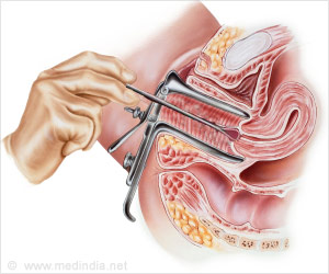
The doctor uses a speculum to open the vaginal canal and visualize the cervix. He then inserts a thin plastic or metal tubular device through the cervix into the uterus to remove a tiny piece of the inner lining tissue.
Usually no anesthesia is required, but taking Ibuprofen 30-60 minutes prior to the procedure can help reduce cramping and pain.
In some cases, a small amount of lidocaine anesthetic may be inserted into the uterine cavity to minimize discomfort.
What are the risks of endometrial biopsy?
There are very few risks with endometrial biopsy. The leading risk is pain or cramping, but this typically subsides following the procedure. Other less common risks are feeling faint, possible infection and rarely perforation of the uterus.


